Today, I was cleaning the house. I used the cleaning spray called Kaboom and did opposite of what the bottle demanded, which was to clean in a well-ventilated area. I'm usually not that careless when I clean but for some reason, I forgot to pop open the window, and well, I felt the affects.
After I finished, I felt like I had a cough that could not come out. And so, being the person I am, I decided to look up the side-effects of inhaling chemical in depth. Of course I know it's not good, but I wanted to see exactly how it effects the respiratory system.
The technical term for this feeling is chemical pneumonitis. The lungs become inflamed when chemical fumes are inhaled and cause difficulty in breathing and may lead to choking. There can both be acute and chronic chemical pneumonitis. Both show symptoms of shortness of breath, cough, and difficulty breathing. The chronic condition may lead to lack of oxygen reaching the body due to difficulty in breathing. which may lead to respiratory failure and death (MedlinePlus, 2014).
Because the respiratory system was a system in focus most recently in class, my unfortunate encounter with chemicals couldn't have come at a more relevant time.
Tuesday, November 24, 2015
Monday, November 23, 2015
Investigations: Journal Post 4
In class, we examine slides that are often prepared with the H&E staining process. While we have gone over H&E stains before, I am choosing to us this weeks blog as a focus on the preparation of these tissue slides in order to refresh myself about the process of H&E stains and how different tissue types respond to this type of staining.
According to a journal entry in Basic Methods of Microscopy, H&E stains have been used to years. They are highly responsive to the different components of body tissue and display them well. Hematoxylin stains stains tissues blue by way of staining nucleic acids while eosin stains tissues pink by way of staining proteins. The journal entry was very detailed with how the staining process occurs and how each chemical reacts with the tissue.
My main focus during investigation and research of H&E stains was to determine why they are more preferred over other methods of staining. Based on LabCE, H&E stains are most common because they provide more depth to an image. The The structures can be differentiated from one another based on the different colors that come from the stains. This helps those studying the tissues determine whether it is abnormal tissue or not. The entry where this question was found was very straightforward with stating why H&E stains are more effective, however, it only mentioned how it is beneficial in pathology.
Thursday, November 5, 2015
Definitions: Journal Post 10
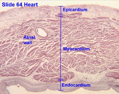
Endocardium- squamous endothelial cells,
In this journal post, I wanted to focus on the cardiovascular system and provide myself another resource for understanding blood flow and how it moves through this system. I also wanted to define and give a more thorough explanation of certain terms.
Circulatory Pattern:
Heart ➢ Artery ➢ Arteriole ➢ Capillary ➢ Venule ➢Vein ➢ Heart

Endocardium- squamous endothelial cells, connective tissue, smooth muscle cells; This layer of the heart provide a smooth surface for blood contraction and pumping. It is the innermost lining of the heart (Boundless).
Myocardium- cardiac muscle cells; This is the muscle tissue of the heart. Cardiac muscle cells are found here and allow for contraction and conductivity of electricity, which need needed for contraction. The cells include intercalated discs which hold cells together for contraction (Boundless).
Epicardium- mesothelium, veins and nerves that supple to heart; This layer of tissue is a layer of connective tissue and serves as a layer of protection for the heart underneath of pericardium (Boundless).


Sources:
https://www.boundless.com/physiology/textbooks/boundless-anatomy-and-physiology-textbook/the-cardiovascular-system-18/the-heart-172/layers-of-the-heart-walls-864-636/
In this journal post, I wanted to focus on the cardiovascular system and provide myself another resource for understanding blood flow and how it moves through this system. I also wanted to define and give a more thorough explanation of certain terms.
Circulatory Pattern:
Heart ➢ Artery ➢ Arteriole ➢ Capillary ➢ Venule ➢Vein ➢ Heart
Endocardium- squamous endothelial cells, connective tissue, smooth muscle cells; This layer of the heart provide a smooth surface for blood contraction and pumping. It is the innermost lining of the heart (Boundless).
Myocardium- cardiac muscle cells; This is the muscle tissue of the heart. Cardiac muscle cells are found here and allow for contraction and conductivity of electricity, which need needed for contraction. The cells include intercalated discs which hold cells together for contraction (Boundless).
Epicardium- mesothelium, veins and nerves that supple to heart; This layer of tissue is a layer of connective tissue and serves as a layer of protection for the heart underneath of pericardium (Boundless).


Sources:
https://www.boundless.com/physiology/textbooks/boundless-anatomy-and-physiology-textbook/the-cardiovascular-system-18/the-heart-172/layers-of-the-heart-walls-864-636/
Subscribe to:
Posts (Atom)