This is the final journal post of the semester! Firstly, I'd like to start by saying that I very much enjoyed Histology. The class was so interesting and it covered everything. It hadn't realized how much tissues mattered until I discovered they literally make up our entire bodies. Seems kind of like a no-brainer now that I think about it. Sometimes the extremely obvious doesn't feel so obvious at the moment. Similarly, the very components of our body don't seem so large alone but when you think of it at a much larger scale, it's a bit breath taking. There are an innumerable amount of cells in our body and they all have a unique job. Some grow up to be a skin cell while others become mucous layers. It's incredible to think about how specific our bodies are and their functions. If these components don't work together flawlessly, our bodies wouldn't run correctly and can lead to greater consequences.
Alongside that, I've noticed something about myself. I am constantly referring to thing I've learned. Ask anyone I talk to and I'm sure they can tell you everything about the lymphatic system thanks to me. They may still not have a clue about what a Peyer's patch is but they can definitely tell you what it does and where it is. Talking about it everyday and fitting it into my daily life has definitely helped me remember it and think of it as more than just something I learned in class.
Overall, I am satisfied with how my semester went and am excited to continue to next semester.
Monday, December 7, 2015
Saturday, December 5, 2015
Investigations: Journal Post 12
The most recent weeks, we have been discussing the digestive system in class and mentioned the gallbladder and how it can be removed from the body without leading to major complications. I wanted to take this blog to go more into depth about complications that occur with the gallbladder and why it may need to be removed.
In SAGES article "Laparoscopic Gallbladder Removal (Cholecystectomy) Patient Information from SAGES", an in-depth discussion about gallbladder disease and removal are given. The gallbladder functions in the digestive system by collecting bile which is then used for digestion. The bile travels from the liver to the gallbladder to the small intestines. In some people, gallstones form and when they do, they can lead to some major issues. Gallstones are typically small formations and can be passed through the gallbladder. In some cases, the stones may form very large and can cause a blockage, preventing bile from getting through to the small intestines. The blockage can lead to swelling, which may lead to rupture. Common symptoms are abdominal pain, vomiting, and fever.
A journal known as Gut and Liver, spoke about what some causes of gallbladder disease are. According to Gut and Liver, gallstones can be diagnosed best through the process of ultrasonography. It also mentions that often times, major risk factors are those that cannot be modified. High cholesterol gallstone formations can depend on genetics as well as age, gender, family history, and ethnic background. Overall, the direction that society is headed in regards to health seems to be causing an increase in gallstone cases.
The most effective way to resolve gallstones seems to be surgery. Gallstones can not be treated themselves, although making dietary changes may help. Overall, removal of the gallbladder may be necessary depending on the severity of the case.
Sources:
Society of
American Gastrointestinal and Endoscopic Surgeons. (2015). Laparoscopic
Gallbladder Removal (Cholecystectomy) Patient Information from SAGES. Retrieved
12 5, 2015, from SAGES:
http://www.sages.org/publications/patient-information/patient-information-for-laparoscopic-gallbladder-removal-cholecystectomy-from-sages/
About this Article: This article was an online publishing by an organization that specialized in gastrointestinal medicine. The article seemed a bit biased as it was trying to push for a particular removal of the gallbladder, cholecystectomy. Overall, the article was very informative and seemed to have come from a credible source.
Stinton, L. M., & Shaffer, E. A. (2012). Epidemiology ofGallbladder Disease: Cholelithiasis and Cancer. Gut and Liver , 6(2), 172.
About this Article: This article focused mostly on the diagnosis and causes for gallbladder disease. It did not seem bias or to push for any specific point as it seemed to have hit all points of risk and did not focus on one specifically.
Reflections: Journal Post 11
As the semester comes to a close, it is becoming brutally clear how quickly life can go by. Just a few years ago, I walked through these halls as a first time college freshman with so much hope. Today, I feel like the time spent thus far has been so incredible. I have learned and experienced so much. Sometimes I sit in class clueless but then most times I can look at a subject and think to myself "I ACTUALLY KNOW THIS!" Often, information from other classes creeps in and it's all a giant circle of information twisted all together.
The semester is nearing its end and for the first time in a very long time, I'm not nervous or anxious about proceeding with my career. Before I would feel unprepared but I will end this semester feeling determined and empowered thanks to all the wonderful professors and students I've met along the way.
The semester is nearing its end and for the first time in a very long time, I'm not nervous or anxious about proceeding with my career. Before I would feel unprepared but I will end this semester feeling determined and empowered thanks to all the wonderful professors and students I've met along the way.
Thursday, December 3, 2015
Definitions: Journal Week 14
This week, focus was on the digestive systems and with an exam coming in close, I wanted to use this blogpost to showcase the major organs in digestive system II and III as well as their structures.
Alimentary Canal:
Functions:
1) Physical barrier to noxious substances, antigens, pathogenic organisms.
2) Immune function (ring of lymphatic tissue surrounding the oral cavity).
3) Secretion (e.g digestive enzymes, mucin, HCl)
4) Absorption


Alimentary Canal:
Functions:
1) Physical barrier to noxious substances, antigens, pathogenic organisms.
2) Immune function (ring of lymphatic tissue surrounding the oral cavity).
3) Secretion (e.g digestive enzymes, mucin, HCl)
4) Absorption
Mucosa: Epithelium
Submucosa: Irregular connective tissue
Muscularis externa: Layer of muscle tissue, contracts and assists with movement down the canal.

Liver: functions in processing and storing nutrients
-Produces albumin, glycoprotein
Exocrine function: secretes bile
Endocrine function: modifies hormones
-Thyroxine
-Vitamin D, A, K

Pancreas: endocrine and exocrine functions
Exocrine functions: produced by acini cells.
-peptidases
-sodium bicarbonate
Endocrine functions:
-Insulin
-Glucagon

Tuesday, November 24, 2015
Encounters: Journal Post 7
Today, I was cleaning the house. I used the cleaning spray called Kaboom and did opposite of what the bottle demanded, which was to clean in a well-ventilated area. I'm usually not that careless when I clean but for some reason, I forgot to pop open the window, and well, I felt the affects.
After I finished, I felt like I had a cough that could not come out. And so, being the person I am, I decided to look up the side-effects of inhaling chemical in depth. Of course I know it's not good, but I wanted to see exactly how it effects the respiratory system.
The technical term for this feeling is chemical pneumonitis. The lungs become inflamed when chemical fumes are inhaled and cause difficulty in breathing and may lead to choking. There can both be acute and chronic chemical pneumonitis. Both show symptoms of shortness of breath, cough, and difficulty breathing. The chronic condition may lead to lack of oxygen reaching the body due to difficulty in breathing. which may lead to respiratory failure and death (MedlinePlus, 2014).
Because the respiratory system was a system in focus most recently in class, my unfortunate encounter with chemicals couldn't have come at a more relevant time.
Monday, November 23, 2015
Investigations: Journal Post 4
In class, we examine slides that are often prepared with the H&E staining process. While we have gone over H&E stains before, I am choosing to us this weeks blog as a focus on the preparation of these tissue slides in order to refresh myself about the process of H&E stains and how different tissue types respond to this type of staining.
According to a journal entry in Basic Methods of Microscopy, H&E stains have been used to years. They are highly responsive to the different components of body tissue and display them well. Hematoxylin stains stains tissues blue by way of staining nucleic acids while eosin stains tissues pink by way of staining proteins. The journal entry was very detailed with how the staining process occurs and how each chemical reacts with the tissue.
My main focus during investigation and research of H&E stains was to determine why they are more preferred over other methods of staining. Based on LabCE, H&E stains are most common because they provide more depth to an image. The The structures can be differentiated from one another based on the different colors that come from the stains. This helps those studying the tissues determine whether it is abnormal tissue or not. The entry where this question was found was very straightforward with stating why H&E stains are more effective, however, it only mentioned how it is beneficial in pathology.
Thursday, November 5, 2015
Definitions: Journal Post 10
Endocardium- squamous endothelial cells,
In this journal post, I wanted to focus on the cardiovascular system and provide myself another resource for understanding blood flow and how it moves through this system. I also wanted to define and give a more thorough explanation of certain terms.
Circulatory Pattern:
Heart ➢ Artery ➢ Arteriole ➢ Capillary ➢ Venule ➢Vein ➢ Heart

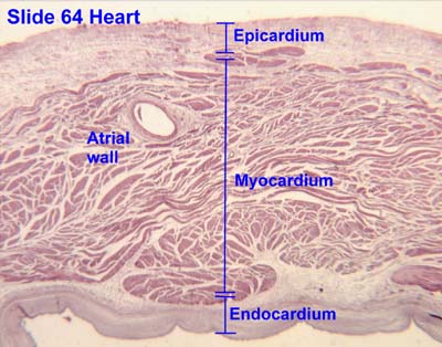
Endocardium- squamous endothelial cells, connective tissue, smooth muscle cells; This layer of the heart provide a smooth surface for blood contraction and pumping. It is the innermost lining of the heart (Boundless).
Myocardium- cardiac muscle cells; This is the muscle tissue of the heart. Cardiac muscle cells are found here and allow for contraction and conductivity of electricity, which need needed for contraction. The cells include intercalated discs which hold cells together for contraction (Boundless).
Epicardium- mesothelium, veins and nerves that supple to heart; This layer of tissue is a layer of connective tissue and serves as a layer of protection for the heart underneath of pericardium (Boundless).


Sources:
https://www.boundless.com/physiology/textbooks/boundless-anatomy-and-physiology-textbook/the-cardiovascular-system-18/the-heart-172/layers-of-the-heart-walls-864-636/
In this journal post, I wanted to focus on the cardiovascular system and provide myself another resource for understanding blood flow and how it moves through this system. I also wanted to define and give a more thorough explanation of certain terms.
Circulatory Pattern:
Heart ➢ Artery ➢ Arteriole ➢ Capillary ➢ Venule ➢Vein ➢ Heart
Endocardium- squamous endothelial cells, connective tissue, smooth muscle cells; This layer of the heart provide a smooth surface for blood contraction and pumping. It is the innermost lining of the heart (Boundless).
Myocardium- cardiac muscle cells; This is the muscle tissue of the heart. Cardiac muscle cells are found here and allow for contraction and conductivity of electricity, which need needed for contraction. The cells include intercalated discs which hold cells together for contraction (Boundless).
Epicardium- mesothelium, veins and nerves that supple to heart; This layer of tissue is a layer of connective tissue and serves as a layer of protection for the heart underneath of pericardium (Boundless).


Sources:
https://www.boundless.com/physiology/textbooks/boundless-anatomy-and-physiology-textbook/the-cardiovascular-system-18/the-heart-172/layers-of-the-heart-walls-864-636/
Subscribe to:
Posts (Atom)